


Many protozoa live in the human body. Many of them are pathogenic. Our story is about ten of them, the most. The journal is compiled from historical and most recent publications.
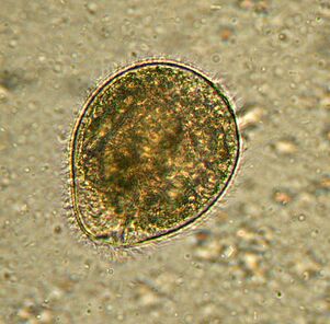
Bigger. BalantidiumBalantidium coli
The largest protozoan is a human parasite, and the only ciliate of this society. Its dimensions vary from 30 to 150 microns in length and 25 to 120 microns in width. For comparison: the length of the malarial plasmodium at the largest stage is about 15 microns, and several times less than that of the balantidium of intestinal cells, among which the ciliates live. An elephant in a china shop.
Distributedwherever there are pigs - its main carriers. Usually lives in the submucosa of the colon, although in humans it also occurs in the pulmonary epithelium. It feeds onB. colibacteria, food particles, fragments of host epithelium. In animals, the infection is asymptomatic. People can develop severe diarrhea with a bloody, viscous discharge (balantidiasis), sometimes ulcers form in the walls of the colon. It is rare to die from balantidiasis, but it causes chronic exhaustion.
People get infected through dirty water or food containing cysts. The infection rate in humans does not exceed 1%, while pigs can be infected anywhere in the world.
treatedwith antibiotics, no reports of drug resistance have yet been reported for this ciliate.
Discoveredby the Swedish scientist Malstem in 1857. Today, balantidiasis is associated with the tropics and subtropics, with poverty and poor hygiene.
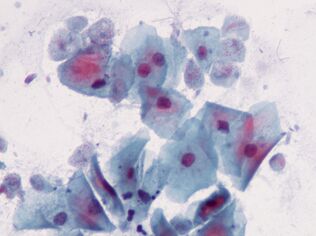
The very first. Oral amoebaEntamoeba gingivalis
The first parasitic amoeba found in humans. The description of amoeba was published in 1849 in the oldest scientific journal. Amoeba is found in dental plaque, hence the name from the Latin gums - gums.
Livesin the mouths of almost everyone with sore teeth or sore gums, inhabit pockets of gums and plaque. It feeds on epithelial cells, leukocytes, microbes and in case of erythrocytes. It is rare in people with a healthy oral cavity.
This small protozoan, 10 to 35 µm in size, does not come out into the environment and does not form cysts, it is transmitted to another host by kissing, dirty dishes or contaminated food. E. gingivalisis considered an exclusively human parasite, but it is sometimes found in captive cats, dogs, horses and monkeys.
At the beginning of the 20th century,E. gingivaliswas described as the causative agent of periodontal disease because it is always present in inflamed dental cells. However, its pathogenicity has not been proven.
Drugsthat affect this amoeba are unknown.
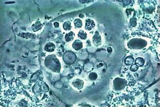
The most common. Amoeba dysenteryEntamoeba histolytica
This intestinal parasite with blood enters the tissues of the liver, lungs, kidneys, brain, heart, spleen, genitals. Eat what he gets: food particles, bacteria, red blood cells, white blood cells, and epithelial cells.
Distributedeverywhere, especially in the tropics. Usually, people get infected by swallowing a cyst.
In temperate countries, the amoeba tends to stay in the lumen of the gut and the infection is asymptomatic. In the tropics and subtropics, the pathological process often begins:E. histolyticaattacks the walls. The reasons for the transition to the pathogenic form are still unclear, but several molecular mechanisms of what is happening have already been described. So, it is clear that amoeba secretes lysing substances, penetrates mucus and kills cells. Apparently, the amoeba can destroy the host cell in two ways: by triggering apoptosis in it or by simply chewing pieces. The first method has long been considered the only one. By the way, the mechanism of cell suicide at record speed - within minutes - has not been identified. The second method was described quite recently, the authors called it trogocytosis from the Greek "three" - gnawing. It should be noted that the amoeba which stings the cells abandon their prey as soon as it dies. Others can completely phagocytose dead cells. It is assumed that biting and devouring cells differ in pattern of gene expression.
Now the ability of the amoeba to enter the bloodstream, liver and other organs is associated with trogocytosis.
Amebiasis is a fatal disease, around 100, 000 people die each year from infection withE. histolytica.
The dysenteric amoeba has a non-pathogenic twin,E. dispar, so microscopy is not enough to diagnose the disease.
To healshould be destroyed as mobileE. histolyticaand cysts.
DescribedE. histolyticaand determined its pathogenic nature in 1875 in a patient with diarrhea. The Latin name for the amoeba was given in 1903 by the German zoologist Fritz Schaudin.Histolyticameans tissue destroyer. In 1906, the scientist died of an amoebic intestinal abscess.
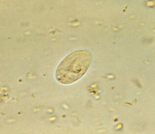
The most common. Intestinal lambliaGiardia lamblia (G. intestinalis)
Giardia, the most common intestinal parasite, is ubiquitous. 3-7% of people in developed countries and 20-30% of developing countries are infected. This represents around 300 million people.
Parasites livein the duodenum and bile ducts of the host, where they float, working with flagella, and then attach to the epithelium using a disksticky located under the cell. On 1 cm2, the epithelium sticks to a million lamblia. They damage the villi, which interferes with the absorption of nutrients, causing inflammation of the mucous membranes and diarrhea. If the disease affects the bile ducts, it is accompanied by jaundice.
Giardiasis is a disease of dirty hands, water and food. The life cycle of the protozoan is simple: in the intestine there is an active form, and at the exit of fecal masses there are stable cysts. To become infected, it is enough to swallow a dozen cysts, which in the intestines will again turn into an active form.
The main secretof the ubiquity of lamblia in the variability of surface proteins. The human body fights antibodies to lamblia and, in principle, is able to develop immunity. But people living in the same area and drinking the same water are infected again and again by the descendants of their own parasites. Why? Because during the transition from the active phase to the cyst and vice versa, lamblia changes the proteins against which antibodies are produced - variant-specific surface proteins. There are about 190 variants of these proteins in the genome, but only one is always present on the surface of an individual parasite; translation of others is interrupted by the RNA interference mechanism. And the change happens about once every ten generations.
It is treatedwith an antiprotozoal agent with antibacterial activity. The disease goes away within a week, but if the bile ducts become infected, relapses are possible for many years. Cysts are fought by iodizing the water.
DiscoveryGiardia lambliain 1859 by the Czech scientist Vilém Lambl. Since then, the simpler has changed several names, and the current one was received in honor of the French discoverer and parasitologist Alfred Giar, who did not describe lamblia.
And the first sketch of Giardia was made by Anthony van Leeuwenhoek, who found it in his own upset chair. It was in 1681.
By the way, Giardia is also evolutionarily very old, almost straight from the ancestor of all eukaryotes.
The most intimate. Trichomonas vaginalisTrichomonas vaginalis.
The simplest, which is sexually transmitted. It lives in the vagina and in men - in the urethra, epididymis and prostate, it is transmitted sexually or through wet washcloths. Babies can get infected through the birth canal.T. vaginalishas 4 flagella at the anterior end and a relatively short wavy membrane; if necessary, it releases pseudopodia. The maximum size of Trichomonas is 32 by 12 microns.
Trichomonas is moreprevalentthan the causative agents of chlamydia, gonorrhea and syphilis combined. It affects about 10% of women, and possibly more, and 1% of men. The latter figure is unreliable because it is more difficult to detect the parasite in humans.
T. vaginalisfeeds on microorganisms, including lactic acid bacteria in the vaginal microflora, which maintain an acidic environment, and thus create an optimum pH for itself above4, 9.
Trichomonas destroys mucous cells, causing inflammation. About 15% of infected women complain of symptoms.
It is treatedwith an antibacterial medicine. As a preventive measure, a regular shower with diluted vinegar is recommended.
Describedin 1836 by the French bacteriologist Alfred Donne. The scientist did not understand that there was a pathogenic parasite in front of him, but he determined the size, appearance and type of movement of the simpler.
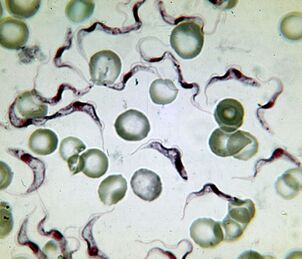
The deadliest. The causative agent of sleeping sicknessTrypanosoma brucei
The causative agent of African sleeping sickness is the deadliest protozoan. An infected person dies without treatment. The trypanosome is an elongated flagellate 15–40 µm long. There are two subspecies which cannot be distinguished externally. Disease caused byT. brucei gambiense, lasts 2 to 4 years.T. brucei rhodesienseis a more virulent and transient pathogen from which they die after a few months or weeks.
Distributedin Africa, between the 15th parallels of the southern and northern hemispheres, in the natural range of the carrier - blood-sucking insects of the genusGlossina(tsetse flytse). Of the 31 species of flies, 11 are dangerous to humans. Sleeping sickness affects the population of 37 countries south of the Sahara at 9 million km2. Up to 20, 000 people fall ill each year. Today, there are around 500, 000 patients, 60 million of whom live at risk.
From the gut of the flyT. bruceienters the human bloodstream, from there it enters the cerebrospinal fluid and affects the nervous system. The disease begins with fever and inflammation of the lymph nodes, followed by lethargy, drowsiness, muscle paralysis, exhaustion, and irreversible coma.
The lethality of the parasite is associated with its ability to cross the blood-brain barrier. The molecular mechanisms are not fully understood, but it is known that when it enters the brain, the parasite secretes cysteine proteases and also uses certain proteins from the host. In the central nervous system, on the other hand, the trypanosome is immune to immune factors.
The first description of sleeping sickness in the upper part of Niger was left by the Arab scholar Ibn Khaldun (1332-1406). By the beginning of the 19th century, Europeans were already well aware of the initial sign of the disease - swelling of the lymph nodes at the back of the neck (a symptom of Winterbottom), and slave traders paid special attention to this.
DiscoveredT. bruceiScottish microbiologist David Bruce, after whom it is named, and in 1903 he first established the link between thetrypanosome, tsetse fly and sleeping sickness.
Treatmentdepends on the stage of the disease and the drugs cause serious side effects. The parasite has high antigenic variability, so it is impossible to create a vaccine.
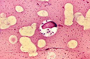
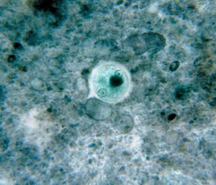
The most extravagant. LeishmaniaLeishmania donovani
Leishmanias have earned the title of the most extravagant parasites, because they live and reproduce in macrophages - cells designed to destroy parasites.L. donovaniis the most dangerous of them. It causes visceral leishmaniasis, colloquially dumdum fever, or kala azar, in which almost all patients die without treatment. But the survivors gain long-term immunity.
There are three subspecies of the parasite.L. donovani infantum(Mediterranean and Central Asia) mainly affects children, dogs are often the reservoir.L. donovani donovani(India and Bangladesh) is dangerous for adults and the elderly, has no natural reservoirs. The AmericanL. donovani chagasi(Central and South America) can live in the blood of dogs.
L. donovani- flagellated no more than 6 microns in length. People get infected after being bitten by mosquitoes of the genusPhlebotomus, sometimes through sexual contact, from babies - passing through the birth canal. Once in the blood,L. donovanienters macrophages, which carry the parasite through internal organs. Reproducing in macrophages, the parasite destroys them. The molecular mechanism of survival in macrophages is quite complex.
Symptoms of the disease- fever, enlarged liver and spleen, anemia and leukopenia, which contribute to secondary bacterial infection. Each year, 500, 000 people fall ill with visceral leishmaniasis and approximately 40, 000 die.
Treatmentheavy - intravenous administration of antimony preparations and blood transfusion.
Taxonomic membershipL. donovaniwas determined in 1903 by the renowned malaria researcher and Nobel Prize winner Ronald Ross. It owes its generic name to William Leishman, and the specific name to Charles Donovan, who in the same 1903 independently discovered protozoan cells in the spleen of deceased kala azar patients, one in London, the other in Madras.
The most difficult lifecycle.Babesia spp.
Babesias, in addition to multistage asexual reproduction in mammalian erythrocytes and sex mites in the intestines of the genusIxodes, have complicated their development through transovarian transmission. From the intestines of a female mite, protozoan sporozoites enter the ovaries and infect the embryos. When the mite larvae hatch, the babesias pass through their salivary glands and, from the first bite, enter the blood of the vertebrate.
DistributedBabesia in America, Europe and Asia. Their natural reservoir is made up of rodents, dogs and cattle. A person is infected with several types:B. microti, B. divergens, B. duncaniandB. venatorum.
The symptoms of babesiosis are similar to malaria - relapsing fever, hemolytic anemia, enlarged spleen and liver. Most people recover on their own, but babesiosis is fatal in patients with weakened immune systems.
TheMethods of treatmentare still under development, while antibiotics are prescribed and, in severe cases, blood transfusions.
Babesia was described by Romanian microbiologist Victor Babes (1888), who discovered it in sick cows and sheep. He decided he was dealing with a pathogenic bacteria which he namedHaematococcus bovis. Babesia has long been considered an animal pathogen, until its discovery in 1957 in a Yugoslav shepherd who died of infection with B. divergens.
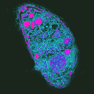
The most influential. The causative agent of toxoplasmosisToxoplasma gondii
T. gondiiis the most powerful parasite because it controls the behavior of intermediate hosts.
Distributedeverywhere, unevenly distributed. In France, for example, 84% of the population is infected, in the UK - 22%.
The life cycle of Toxoplasma consists of two stages: asexual occurs in the body of all warm blood, sexual reproduction is possible only in the epithelial cells of the intestine of the cat. AtT. gondiicould end development, the cat must eat an infected rodent. By increasing the likelihood of this event,T. gondiiblocks the rodent's natural fear of cat urine odor and makes it attractive by targeting a group of neurons in the amygdala. How she does this is unknown. One of the supposed mechanisms of action is a local immune response to infection. It changes the levels of cytokines, which in turn increases the levels of neuromodulators such as dopamine. Toxoplasma also affects human behavior, which manifests itself even at the population level. Thus, in countries with a high level of toxoplasmosis, neuroticism and the desire to avoid uncertainty, new situations are more frequent. It is possible that infection withT. gondiicould lead to cultural changes.
Infectionin humans is often asymptomatic, but with weakened immunity it destroys cells in the liver, lungs, brain, retina, causing acute or chronic toxoplasmosis. The course of the infection depends on the virulence of the strain, the state of the host's immune system and its age - older people are less susceptible toT. gondii.
Treattoxoplasmosis with antiprotozoal drugs.
Describedin 1908 in desert rodents. This honor belongs to the staff of the Institut Pasteur de Tunisie, Charles Nicolas and Luis Manso.
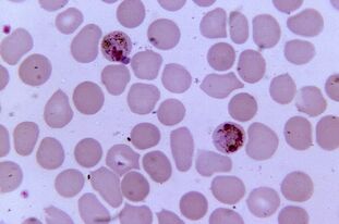
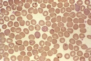
The most pathogenic. PlasmodiumPlasmodium spp.
Plasmodium malaria is the most pathogenic parasite in humans. The number of patients with malaria can reach 300-500 million, and the death rate during epidemics - 2 million. The disease still claims three times as many lives as armed conflict.
Five types of Plasmodium cause malaria in humans:Plasmodium vivax, P. falciparum, P. malariae, P. ovaleandP. knowlesi, which affectsalso macaques.
Distributedin the range of vectors - mosquitoesAnopheles, which require a temperature of 16 to 34 ° C and a relative humidity of more than 60%.
Comparison of the genome of the most virulent of the plasmodia,P. falciparum, with the gorilla plasmodia suggests that man was infected by his ancestor of these monkeys. The emergence of this form of Plasmodium is associated with the emergence of agriculture in Africa, which has led to an increase in population density and the development of irrigation systems.
Sexual reproduction of plasmodia occurs in the intestines of mosquitoes, and in the human body it is an intracellular parasite that lives and reproduces in hepatocytes and erythrocytes until the cells burst. 1 ml of the patient's blood contains 1 to 50, 000 parasites.
The disease is manifested by inflammation, periodic fever and anemia, in pregnancy it is dangerous for the mother and the fetus. Erythrocytes infected withP. falciparumobstruct capillaries and, in severe cases, ischemia of internal organs and tissues develops.
TheTreatmentrequires a combination of several drugs and depends on the specific pathogen. Plasmodia become resistant to drugs.